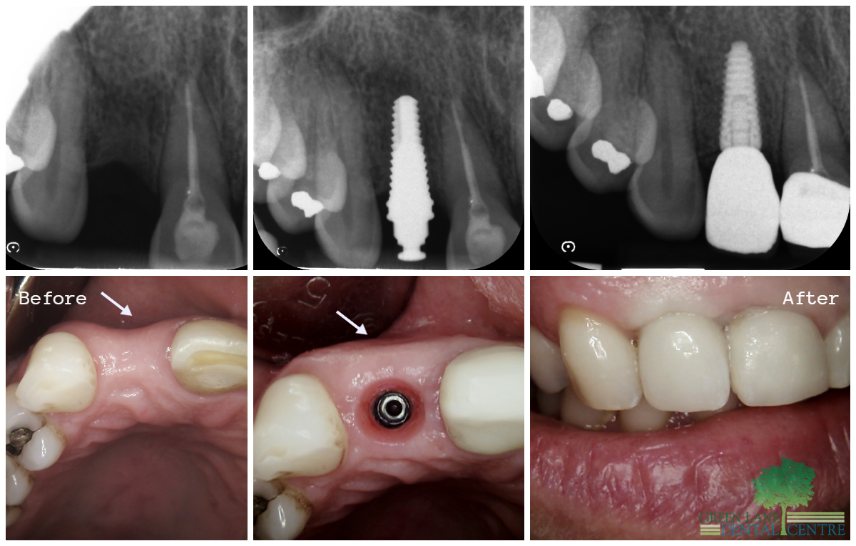
A remarkable aesthetic outcome was achieved in the upper right second tooth area through the replacement with a dental implant.
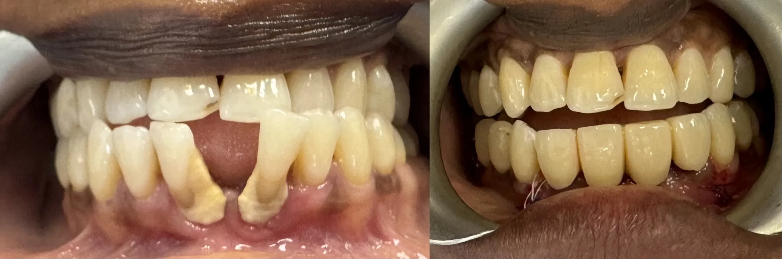
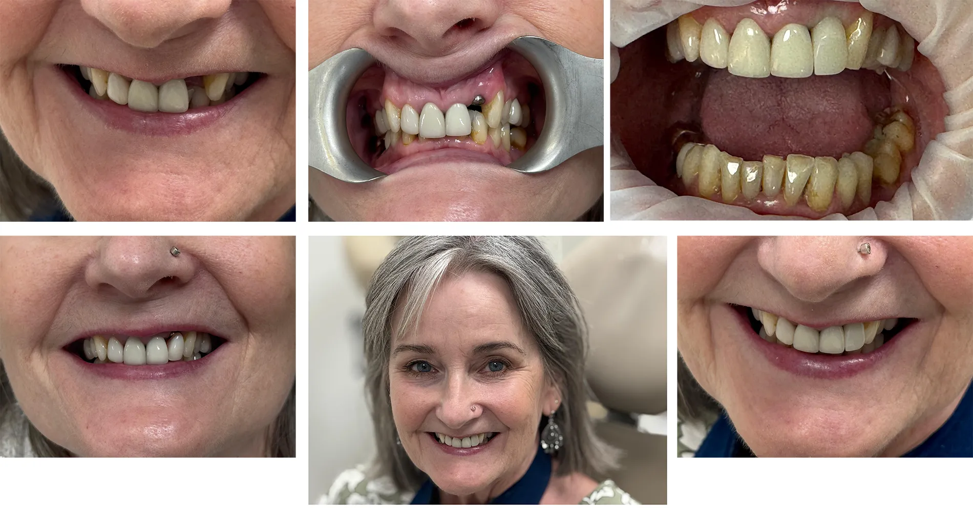
She lost 2 teeth due to gum disease and the other 2 getting loose. Then Extraction 2 lower teeth due to gum disease and place 2 implant then 4 units bridge
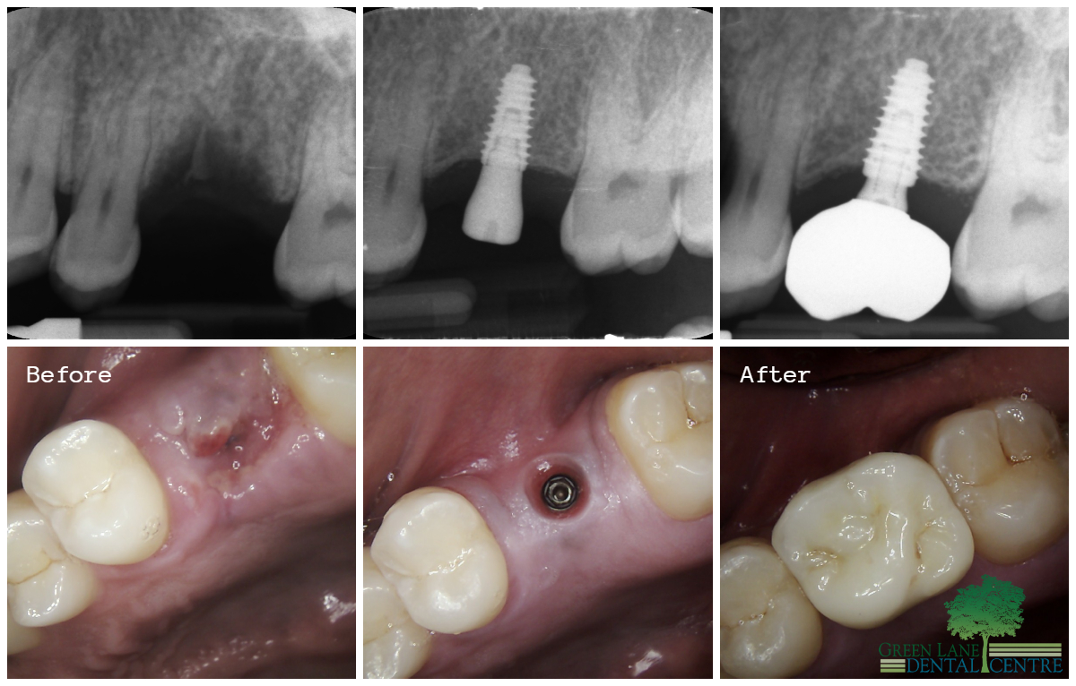
A sinus lift procedure was performed to elevate the sinus membrane, creating space to accommodate a sufficiently long implant for the missing upper right second premolar.
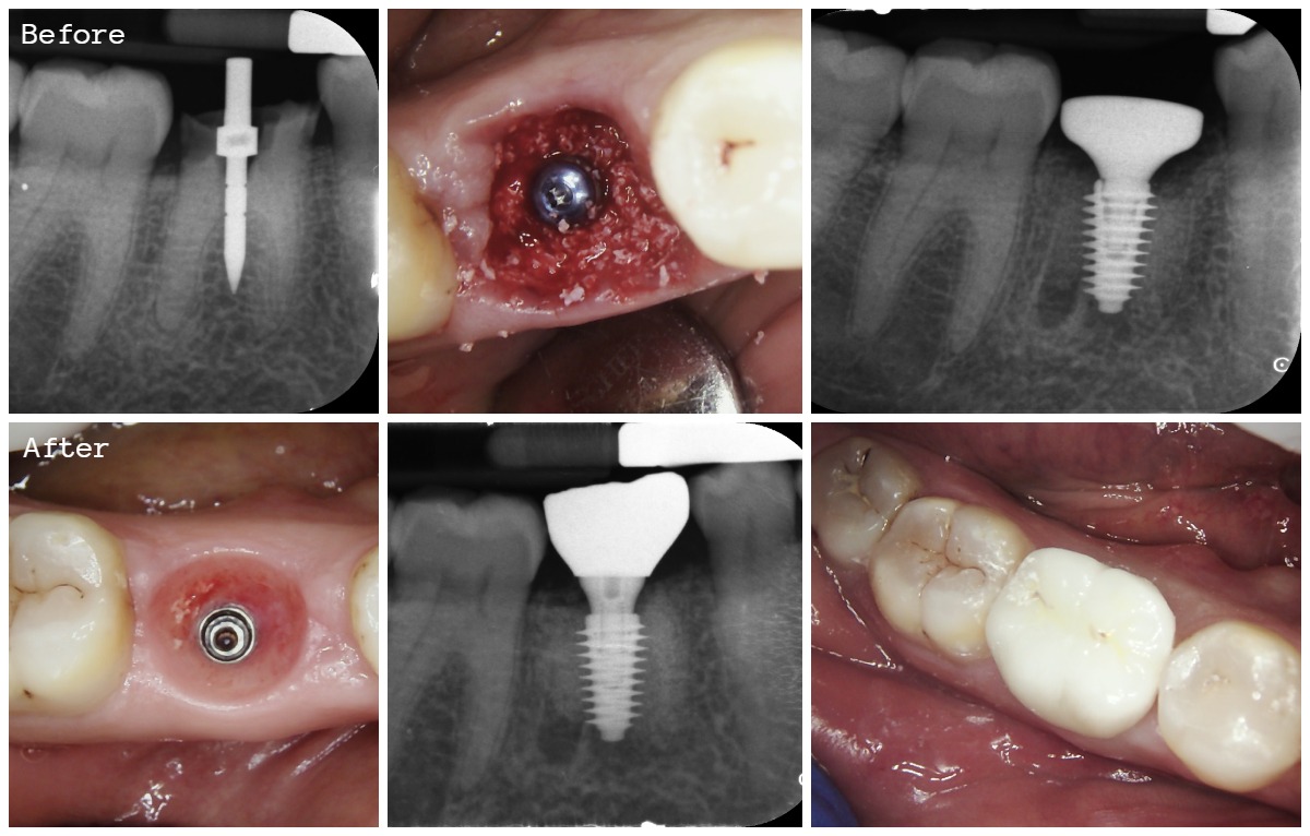
Immediate implant placement with applying bone graft inside the extraction socket
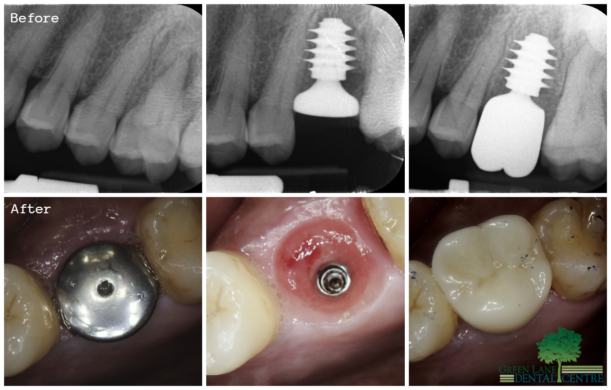
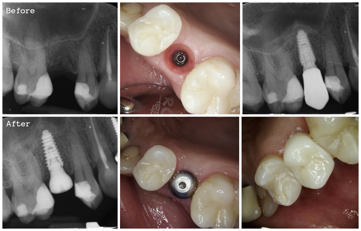
An immediate implant placement procedure was performed for the upper left molar, followed by a remarkable gum healing process over the course of four months. Subsequently, a crown was fabricated and connected to the implant, resulting in a fully restored and functional tooth.
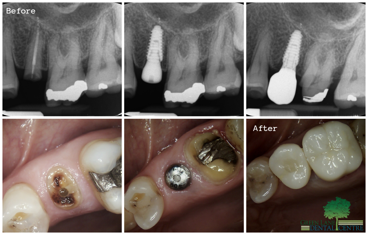
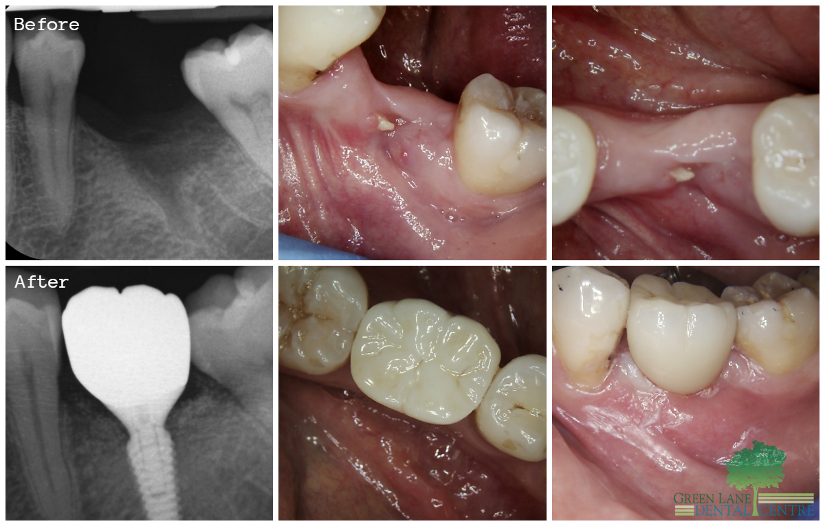
Immediate implant placement was performed for the broken upper left second premolar tooth. After a period of five months, the gum healed beautifully. Subsequently, a crown was fabricated for the implant, and a full porcelain crown was placed on the adjacent tooth, which had a large defective metal filling with a deep fracture.
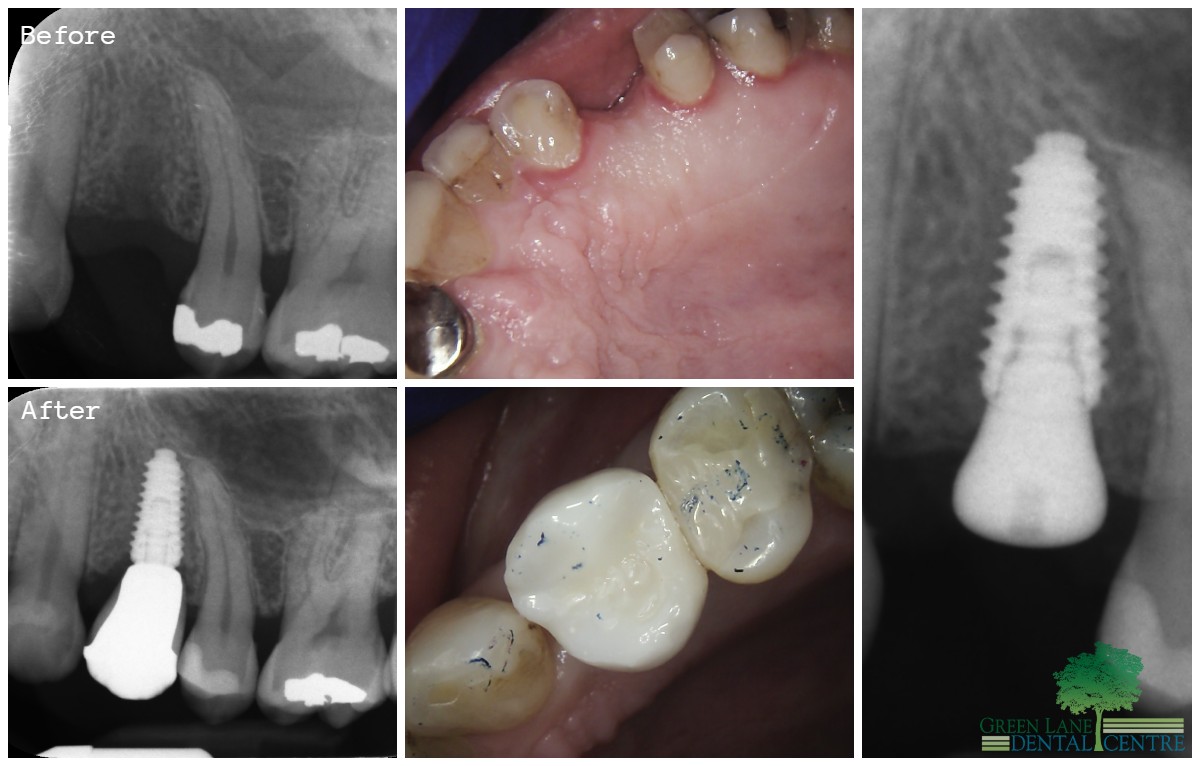
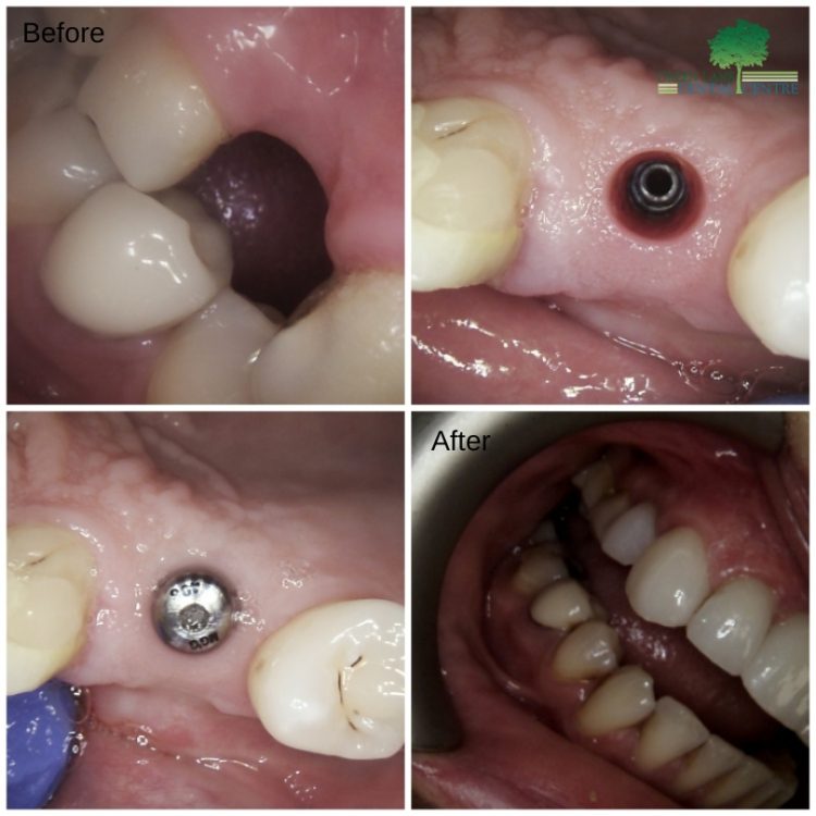
The missing upper right canine in the aesthetic zone was replaced with a dental implant. Simultaneously, a healing cap was connected to the implant to facilitate gum formation. After a period of five months, the crown was fabricated and connected to the implant, completing the restoration process.
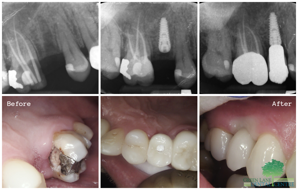
The upper right first premolar was extracted due to deep decay, which also led to the exposure of the nerve on the adjacent tooth as a result of decay under a defective filling. To address this issue, an implant was placed to replace the missing premolar. Additionally, root canal treatment was performed on the upper right first molar. Subsequently, crowns were fabricated for both the implant and the root canal-treated tooth, resulting in a remarkable outcome.
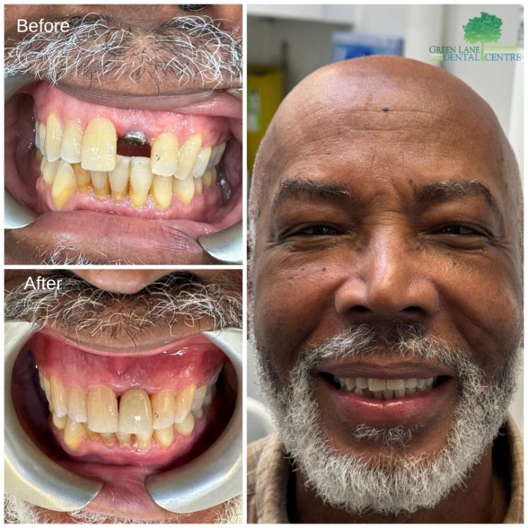
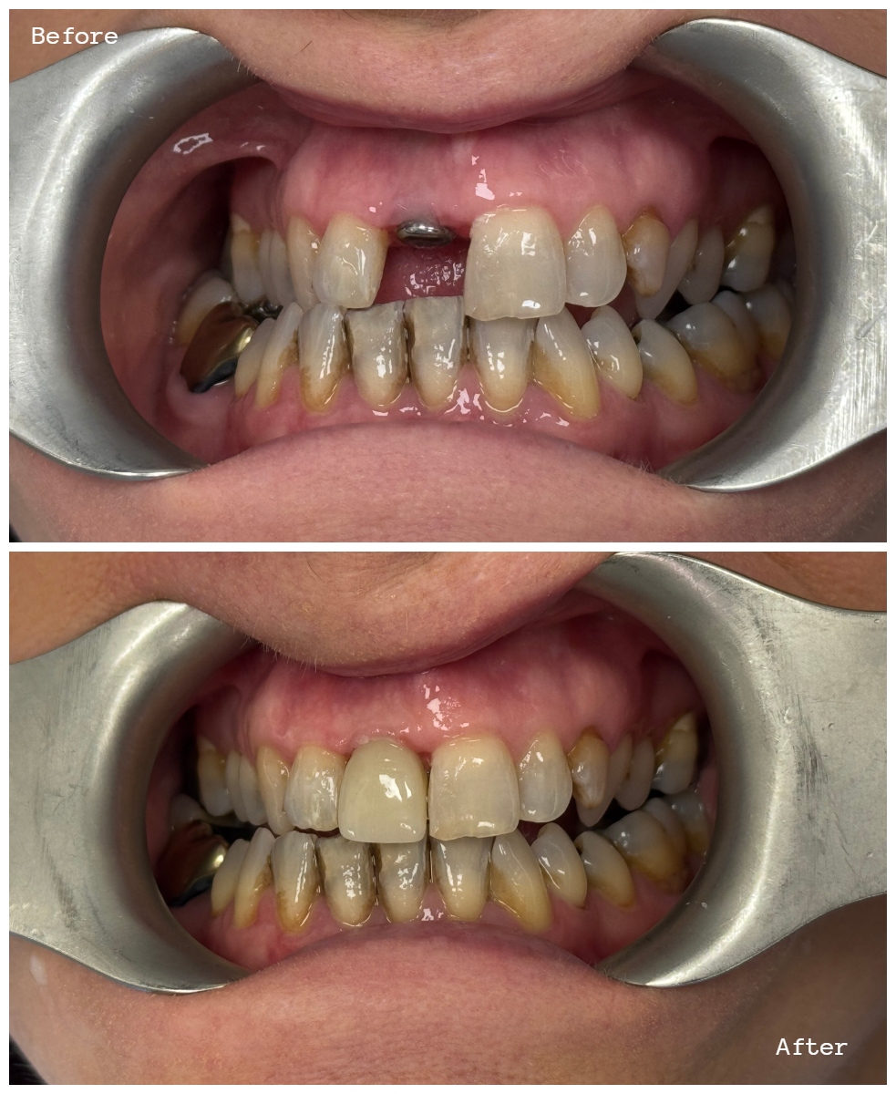
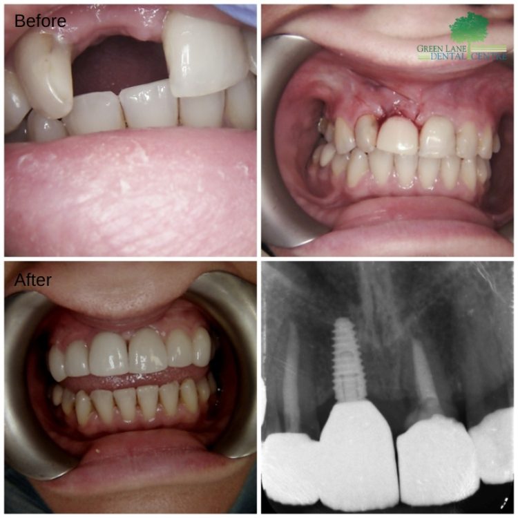
Single implant placement will be performed under local anesthesia, followed by the immediate placement of a temporary bridge to close the gap on the same day. After a period of six months, the crown will be connected to the implant to complete the restoration process.
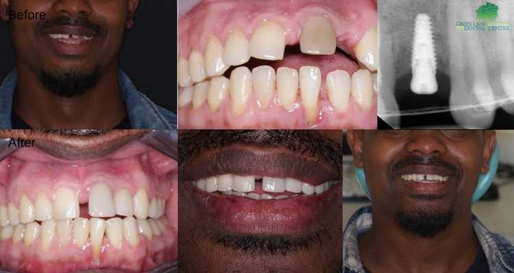
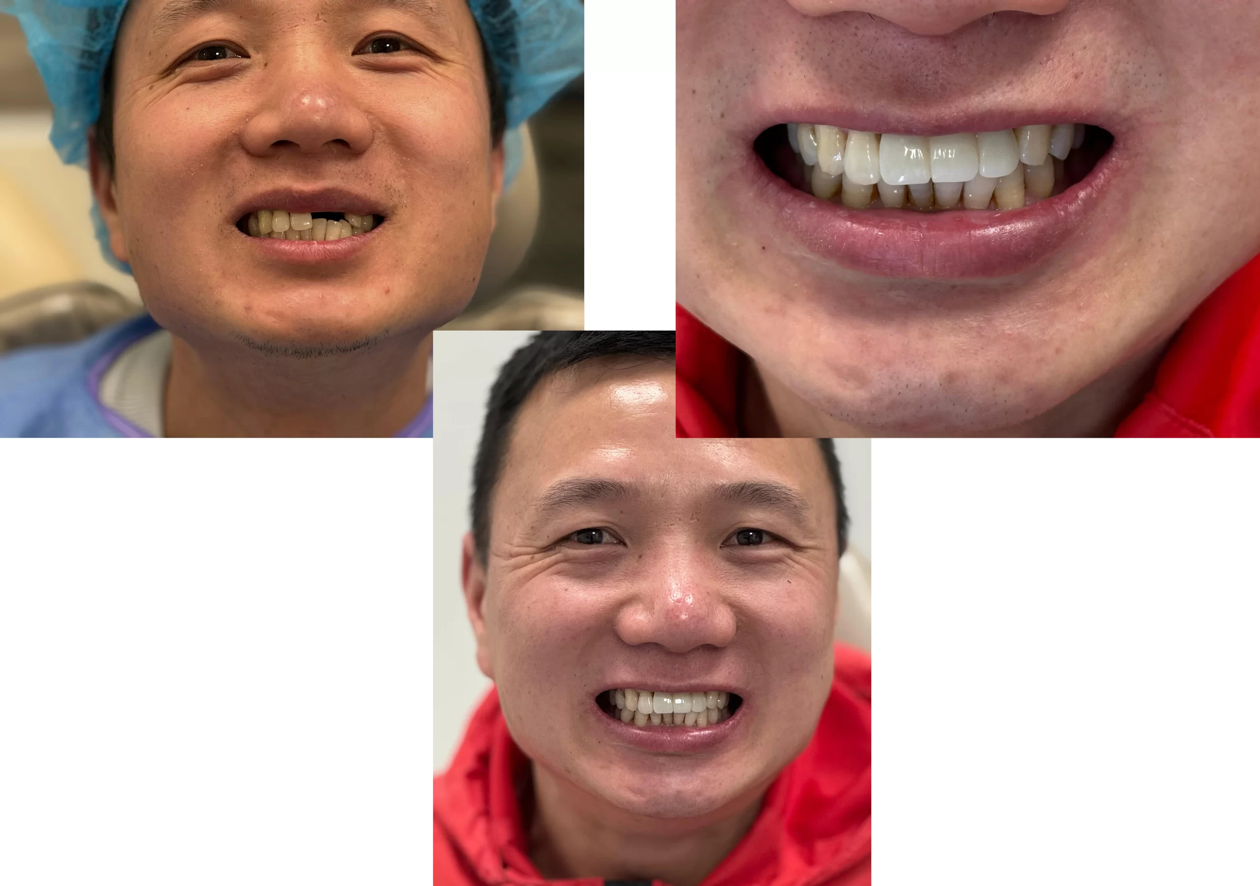
When art and science collaborate, the boundaries of possibility are expanded. This is exemplified in the treatment of a missing Upper Left second tooth, accompanied by a gap between the upper front teeth and outward shifting. The position of the upper left first tooth was meticulously corrected, setting the stage for the restoration of the missing tooth. A single titanium implant, coupled with a metal-free crown, was selected for its compatibility and aesthetic appeal. This approach not only restored the missing tooth but also harmonized the dental alignment and appearance, showcasing the seamless integration of artistic vision and scientific precision in achieving exceptional dental outcomes.
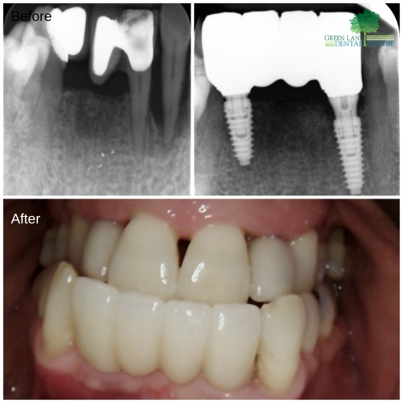
A 4-unit bridge was connected to two implants to replace the lower four front teeth, which were lost due to gum disease.
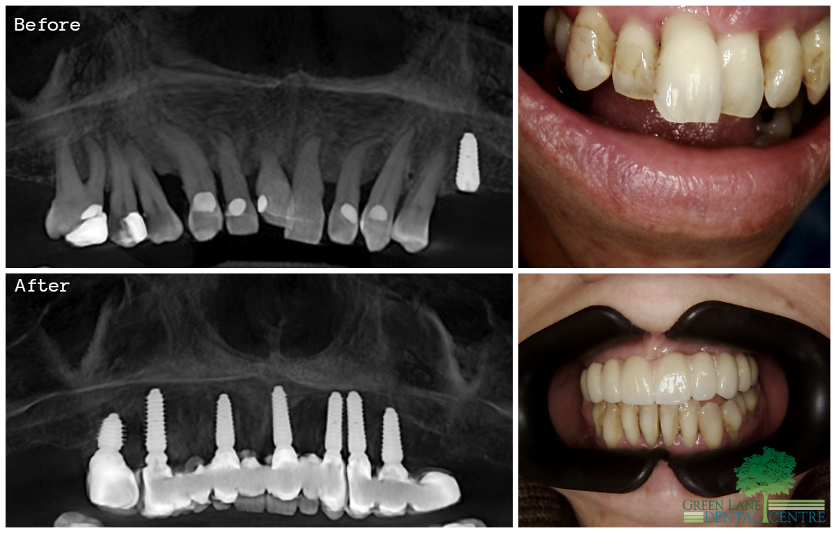
All upper teeth were removed, and seven implants were placed. After five months, all upper teeth were replaced with three different unit bridges. A comprehensive implant placement procedure was conducted for a lady experiencing gum disease, which had led to loose and protruding upper teeth. In response, 7 implants were strategically placed to address the issue. Following this, 3 bridges were meticulously fabricated, replacing a total of 12 teeth. This restoration not only restored functionality but also enhanced the aesthetic appeal of her smile.
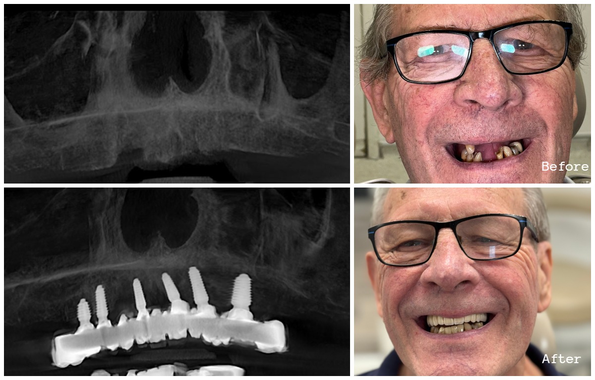
Six implants were placed in the upper jaw of a beloved gentleman, and they will be restored with a full porcelain bridge six months later.
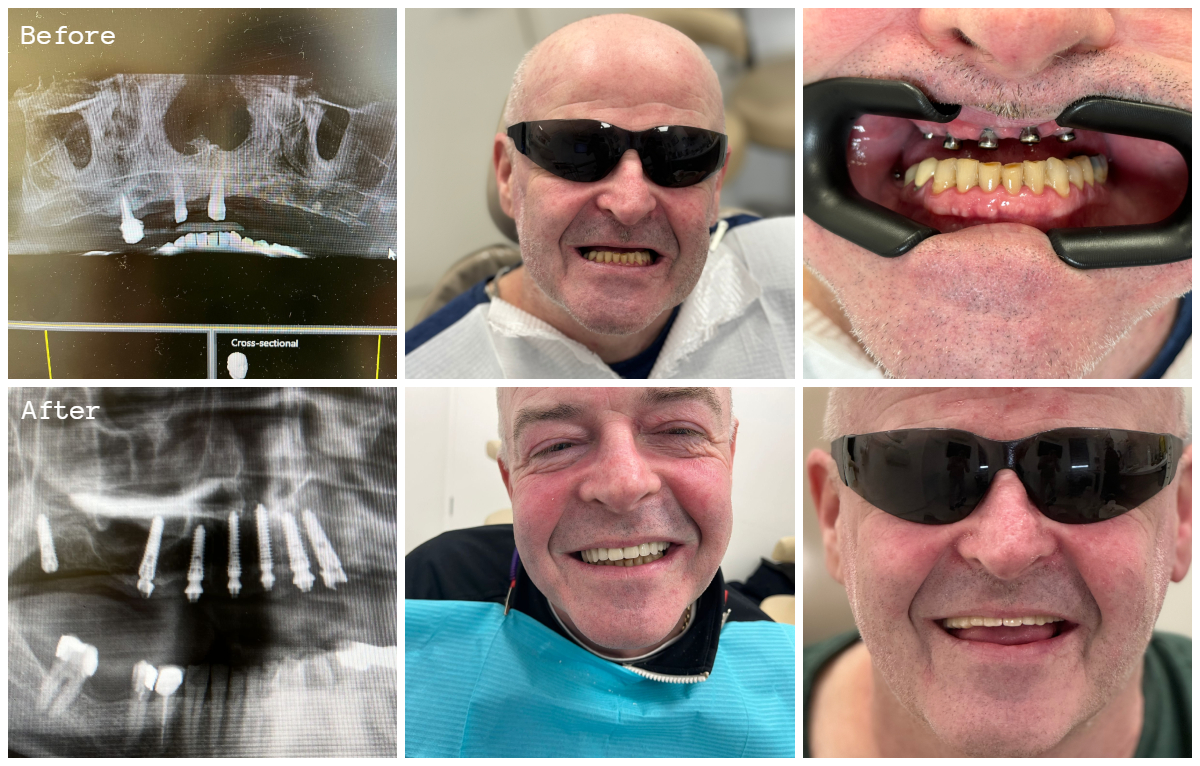
Before and after images show the transition from the initial placement of implants with a temporary bridge to the final full upper arch bridge.
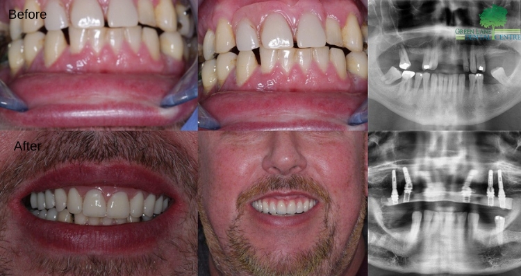
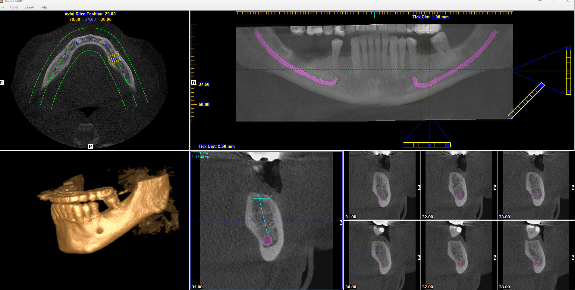
In addition to the lower denture solution, the patient expressed discomfort with his removable upper denture and sought a more permanent solution that could be implemented promptly. Following a thorough consultation, he underwent the extraction of all remaining upper teeth and received six implants (All-on-6) with an immediate fixed bridge loaded the same day. The entire procedure was performed under local anesthesia, guided by a surgical plan informed by his Cone Beam Computed Tomography (CBCT) scan. This fixed bridge serves as a temporary solution while his gums heal from the extractions. He is scheduled to return in six months for the fitting of a permanent bridge, marking the completion of his dental restoration journey.